


Bordetella bronchiseptica in pigs
Bordetella genus belongs to the Beta protebacteria class of which 9 species have been described. Bordetella bronchiseptica is the only important one in pigs and causes atrophic rhinitis...

Respiratory diseases in pigs have a significant economic impact in farm. This is because they directly affect production rates, especially Average Daily Gain (ADG) and, therefore, final weight. Depending on the type of respiratory disease, it can develop asymptomatically or cause a very high number of deaths. Respiratory pathologies have a viral or bacterial origin, but as the first infection progresses, it is common to observe infections caused by different viruses and bacterial.
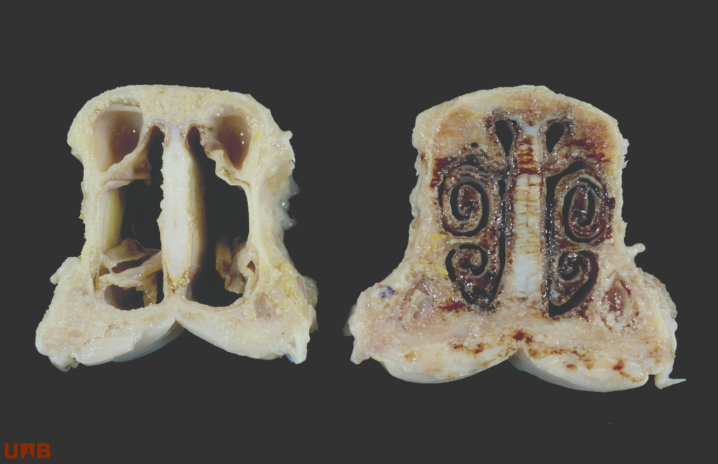
Bordetella genus belongs to the Beta protebacteria class of which 9 species have been described. Bordetella bronchiseptica is the only important one in pigs and causes atrophic rhinitis. Atrophic rhinitis is characterized by lesions in the nasal turbinates and can be presented in two different forms: Non-Progressive Atrophic Rhinitis or Progressive Atrophic Rhinitis (visit link).
brochiseptica has a worldwide distribution and is known to infect poultry and a wide range of wild and domestic mammal species. It has a high prevalence in pigs and is frequently isolated from both pigs with atrophic rhinitis and/or pneumonia and from those that are apparently healthy. It is a gram-negative, aerobic and motile coccobacillus, 1.0 x 0.3 mm in size. It is a non-fermentative bacterium, but positive for oxidase, catalase, urease and citrate. It grows on blood agar and MacConkey agar.
Two strains are distinguished: strains of complex I, which are most frequently isolated from animals, and strains of complex IV, which are most frequently isolated from humans. The bacteria remain viable for up to 45 days in soil and can remain viable for several weeks in water at temperatures between 10 and 37ºC. The half-life in air at room temperature and 75%n relative humidity is 1-2 hours. B. bronchiseptica can be deactivate by sonication, temperature (>60ºC) or formaldehyde treatment.
Virulence factors
Virulence factors are regulated by virulence genes, which require co-expression of the BvgAS system. This system allows the synthesis of toxins, adhesins and other virulence proteins. At temperatures of approximately 25ºC or lower, BvgAS genes are not expressed, and it takes an expression of motility genes, virulence repressed genes (vrg) and genes required for urease production. When the temperature rises, BvgAS genes are expressed and promote colonization of the respiratory tract and cause lesions: first in the upper airways and then in lungs.
The main responsible substances for lesions are: filamentous hemagglutinin (FHA), fimbrial proteins, pertactin, type III secretion system (T3SS), dermonecrotic toxin (DNT), adenylate cyclase toxin (ACT) and tracheal cytotoxin (TCT).
- Filamentous hemagglutinin (FHA). It is a protein associated with the bacterial cell Surface, highly immunogenic and necessary for colonization of the upper respiratory tract.
- Fimbrial proteins. They extend over the entire bacterial surface. They are important for colonization and persistence in the trachea, contribute to the development of biofilms and influence immune responses.
- Pertactin. It contributes to colonization, but its function is still unclear.
- Type III secretion system (T3SS). Immunomodulatory and cytotoxic effects. Contributes to the severity of pneumonic lesions and bacteria’s ability to persistently infect the lungs.
- Dermonecrotic toxin (DNT). It causes bone deformation (turbinate atrophy) and necrosis, haemorrhage, neutrophil accumulation and fibrosis in the lungs.
- Adenylate cyclase toxin (ACT). It alters innate immunoprotective functions, together with FHA and T3SS the target cells are phagocytic cells. In addition, it modulates cytokine production and alters antibody responses.
- Tracheal cytotoxin (TCT). It causes necrosis of the ciliated epithelium that covers the nasal cavity and the conducting airways, resulting in impaired mucociliary clearance function.
The damage caused by these toxins, especially by DNT, facilitates the colonization of other secondary pathogens and cause a greater clinical severity. On fact, secondary infection by Pasteurella mutocida is quate typical and provoke Progressive Atrophic Rhinitis in pigs.
Prevention and Control
B.bronchiseptica locates in the nasal cavity for several months and may persist indefinitely. The introduction of carrier pigs is a source of infection ant the spread within a group is rapid. Therefore, it is important that the animals have a strong immune system, and this is achieved by eliminating or minimizing the predisposing factors: stress, density, hygiene of the facilities, concentration of ammonia in the environment, farm management (all in/all out).
Transmission of bacteria occurs from sows to piglets or between piglets and this transmission is interrupted by vaccination of sows at 6 and 2 weeks before farrowing. Vaccination reduces the severity of the disease and increases piglet performance, although it does not prevent pathogen colonization in piglets. Vaccination of non-immune piglets provides protection against nasal turbinate atrophy, since piglets vaccinated with whole-cell bacterins at 1 and 4 weeks of age develop circulating antibodies that last beyond 12 weeks of age.
The most studied vacines that hace proven most effective are the inactivated ones, which contain whole-cell bacterin, used to control atrophic rhinitis. In addition, their effectiveness is considerably impoved when the vaccine also protects against other pathogens, especially Pasteurella multocida, composed ow whole cells or toxoids of P. multocida. Therefore, the inactivated vaccine against B. bronchiseptica and P. multocida is the best prevention and control measure, reducing the use of antibiotics and improving the productivity and economic performance of the farm.


CATEGORIES
- Antibiotics
- Antioxidants
- Aquaculture
- Deodorizer
- Digestives
- Dissemination
- Enzymes
- Events
- Farm management
- Farm management
- Fats
- Feeding
- Flavorings
- Immunostimulants
- Microbiocide
- Mycotoxins
- Nutritionals
- Pathology
- Pharmacology
- Poultry Farming
- Preservatives
- Pronutrients
- Ruminants
- Swine
- Technology
It may interest you
Viral respiratory diseases in poultry farming: How to prevent them with vaccination systems
Reduction of gases in pig farms: the role of natural additives in emission mitigation
Poultry Nutrition: the role of feed supply systems and their impact on sustainable production
Enterococcus cecorum: A Growing Challenge in Broiler Production

Biosecurity in egg handling: What recommendations to consider?


Leave a Reply
You must be logged in to post a comment.