


Porcine coccidiosis
A frequent pathology that mostly affects lactating piglets

-
- 1 What is porcine coccidiosis?
- 2 What is the economic relevance of porcine coccidiosis?
- 3 Etiology
- 4 Life cycle and transmission of porcine coccidiosis
- 5 Risk factors
- 6 Clinical signs and lesions of porcine coccidiosis
- 7 Diagnosis of porcine coccidiosis
- 8 Treatment and prevention of porcine coccidiosis
What is porcine coccidiosis?
Porcine coccidiosis is a very common parasitic disease of global relevance in porcine farming because of its negative effects on performance and growth of piglets.
The name of this disease is used as a general term to describe clinical signs and lesions caused in pigs by protozoa of the genera Eimeria or Isospora.
These parasites cause tissue damage in the intestine, which alters nutrients absorption and causes diarrhoea and decreased performance.

What is the economic relevance of porcine coccidiosis?
Clinical coccidiosis is one of the main causes of ill-thrift, poor feed conversion and loss of litter uniformity in lactating and post-weaning piglets. Its prevalence is usually higher in countries where pigs are raised intensively (Chart 1).

Etiology
Eimeria and Isospora are genera of coccidia that affect pigs. They are obligate intracellular protozoa that replicate in the intestinal epithelium of the host and cause different degrees of enteritis depending on the species.
Lactating animals are the most prone to suffer this pathology. The vast majority of coccidiosis cases in lactating piglets are produced by Isospora suis, which causes the called “neonatal coccidiosis”. For this reason, Isospora suis is considered the most important coccidia in pigs and is the focus of this article.
Life cycle and transmission of porcine coccidiosis
Transmission is carried out by ingestion of sporulated oocysts by piglets (infective form of the parasite) present in contaminated environment of farrowing barns, that comes either from actual sick piglets or from sick piglets of previous cycles.
In the past, mothers were suspected to be the main source of contamination. However, different studies have revealed that sows shed a low amount of parasites, while the piglet is the main contaminant, shedding around 100,000 oocysts per gram of feces. Mechanical transmission by vectors, such as farm tools or animals, like flies or rodents, may also occur.
Thanks to the digestion process, sporozoites are released from the interior of the oocyst. Then, they penetrate into intestinal cells and perform several cycles of asexual and sexual reproduction, thus destroying the intestinal epithelium and producing a large number of new oocysts. This is a fast cycle that lasts about 3 or 4 days.
Adequate conditions of oxygenation, temperature and humidity are necessary for sporulation. Farrowing barns have ideal environmental conditions to favor sporulation and, therefore, the outbreaks of coccidiosis. These sporulated oocysts are highly resistant and can remain viable for up to a year, which eases the persistence of the infection.
Risk factors
Virulence of each specie and course of the disease are determined by several factors:
Factors that depend on the host
Age, nutrition and immunity of the animal, and existence of concomitant infections that are immunosuppressive or that attack the intestine. Young animals in lactation are the most susceptible to suffer from the disease.
Factors that depend on the parasite
Number of inoculated oocysts and species involved.
External factors
Housing, hygiene and handling conditions.
Clinical signs and lesions of porcine coccidiosis
Intestinal epithelium damage is the source of symptomatology and nutrients absorption alteration which finally leads to weight loss.
The main symptom in lactating piglets suffering from Isospora suis infection is yellowish or grayish diarrhoea (with an acid smell) that leads to dehydration of the animals and considerable weight loss. Signs may appear from the 5th day of life through all the lactation period and even one week after weaning, being more frequent in the second week of life (Figure 1).

It is a disease that courses with high morbidity, although mortality is relatively low if animals receive treatment. Porcine coccidiosis is more severe when combined with secondary agents such as Escherichia coli, Clostridium perfringens or rotavirus, which are frequent in piglets.
Productive parameters are affected for a long time because, after the end of the clinical signs, a period for intestinal villi regeneration is needed.
In post-weaning and fattening, signs are usually less visible, and pasty stools or a subclinical disease with slow growth of the pigs are observed. Generally, adult animals do not show symptoms but are asymptomatic carriers.
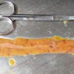
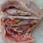
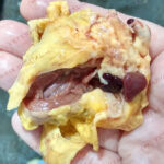
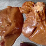
Lesions caused by this disease are visible at the level of the small intestine as jejunitis and ileitis that varies from catarrhal to fibrinonecrotic depending on severity of the infection.

Diagnosis of porcine coccidiosis
Presumptive diagnosis is based on the evaluation of the clinical signs and the poor response to antibiotic treatment.
Definitive diagnosis can be made from a coprology. When coccidiosis is suspected, it is appropriate to sample animals with clinical signs for two or three days to ensure they are shedding oocysts, since, if collection is done in previous or subsequent periods, oocysts may be minimal or even nonexistent. It is important to carry out repeated sampling on the farm in order to detect the parasite.
The most reliable diagnosis is based on identification of the parasite in intestinal sections (histopathology), thanks to the staining of the tissue with Giemsa method.
There are other techniques that ensure the detection of oocysts, such as fluorescence techniques or molecular techniques, but they are not usually used in field conditions.
Differential diagnosis
It is important to differentiate coccidiosis from other enteric processes that show yellowish diarrhoea and appear in the first weeks of life, among which we find colibacillosis or clostridiosis as the most common diseases.
To differentiate them, faeces consistency and blood presence should be considered, but also the age at which diarrhoea outbreak takes place and response to treatment with different drugs. In many cases, pig producers report presence of neonatal diarrhoea, compatible with colibacillosis, which does not respond to antibiotic treatment. This last fact may guide diagnosis towards a parasitic or viral disease, discarding the other options mentioned above.
Treatment and prevention of porcine coccidiosis
It is essential to establish a proper plan of hygiene, disinfection and management of the animals. The system “All in – All out”, separating different productive cycles with a complete emptying of the facilities, has showed beneficial results.
Hygiene between production cycles should include the use of steam and disinfection with ammonium compounds or sodium hypochlorite. Wood or cement surfaces are harder to clean than metal or plastic grill floors. After cleaning, room must be dried correctly prior to entry of the animals.
During the production cycle, periodically checking of the drinking system, removing faeces or changing litter to avoid contamination and to ensure dryness of soil is recommended. In addition, mechanical transmission of oocysts should be avoided by plague control and use different working material (tools and boots) between several pens.
These biosecurity and preventive management measures mentioned above allow to reduce the risk of coccidiosis transmission, but additional measures are always necessary to control the disease, such as the use of drugs or natural products. Vaccines against porcine coccidiosis are not available in the market.
Use of drugs
The drug considered as the most effective for prevention and treatment of porcine coccidiosis is Toltrazuril, active for intracellular stages of the parasite, both asexual and sexual. There are different use guidelines, which include preventive administration in sows during pre and post-farrowing period, or administration in piglets orally or injected.
Resistances development to this drug, in addition to the growing pressure of society on the use of natural alternatives in animal production, has limited its use.
There are products based on natural substances, such as pronutrients, which work as a natural solution to coccidiosis.
Stimulation of immunity by natural products
Products based on pronutrients are a natural solution for the prevention of coccidiosis.
Pronutrients are active molecules derived from plants that induce the expression of genes related to protein synthesis in a target cell. Intestinal optimizer pronutrients promote the activity of the intestinal immune system of piglets. Thanks to their action, the immune system is able to naturally eliminate coccidia. Trophozoites are the most sensitive stage of the parasite to the effects of the immune system, so intestinal optimizers work at this level, causing the elimination of the parasite from enterocytes and avoiding intestinal mucosa damage.
Since intestinal optimizers do not work directly on the pathogen, as they stimulate animal physiology, they do not create resistances and can be administered in continuous programs. In addition, their natural origin assures that no residues on animals are left and no withdrawal period is required.

Bibliography:
- Martínez Moreno, F.J. et al. (2011). Coccidiosis porcina: posibilidades de control. Anaporc: revista de la Asociación de Porcinocultura Científica, ISSN 1697-2147, Vol. 8, Nº 83, 2011, págs. 24-30.
- Pié, J. (2016). Parasitosis porcinas: coccidiosis Neonatal, etiología y epidemiología. Veterinaria digital.
- Pié, J. (2016). Parasitosis porcinas: coccidiosis Neonatal, diagnóstico. Veterinaria digital.
- Pié, J. (2016). Parasitosis porcinas: coccidiosis Neonatal, control y prevención. Veterinaria digital.
- Pié, J. (2016). Parasitosis porcinas: coccidiosis Neonatal, un nuevo enfoque en la lucha contra la coccidiosis porcina. Veterinaria digital.
- Quílez Cinca, J. et al. (2003). Coccidiosis porcina. MG Mundo Ganadero, año 14, Nº 156, págs. 52-56.


Leave a Reply
You must be logged in to post a comment.