


Avian necrotic enteritis
What is necrotic enteritis? What is its economic importance? How is it transmitted? What signs or injuries cause? How to prevent it?

Necrotic enteritis is an important disease in poultry production worldwide because of its negative effects on productivity and the development of birds.
It is an acute enterotoxaemia caused by Clostridium perfringens, characterized by severe necrosis of the intestinal mucosa, which alters nutrient absorption and productivity and may cause the death of the bird.
Economic importance of avian necrotic enteritis
Necrotic enteritis is one of the most common diseases in poultry and causes economic losses worldwide. The ban of antibiotic growth promoters has made it a re-emerging disease.
The economic impact is estimated to be around 6 thousand million dollars per year, because of the mortality of its clinical presentation and, particularly, the decrease in productivity caused by the subclinical form.
Etiology
This disease is related to toxins produced by Clostridium perfringens type A and C. Clostridium perfringens is a gram-positive anaerobic bacterium that is part of the intestinal microbiota. The outbreak of the disease is related to an excessive bacterial growth.
It usually affects 2 to 5-week-old birds, although hens during the start or the peak of laying can also be affected.
Biological cycle and transmission of necrotic enteritis in poultry
Clostridium perfringens is a ubiquitous bacterium, usually transmitted through the feco-oral route, when birds eat contaminated feed, water or litter. For this, it is essential to conduct a microbiological control of feed and water to detect the presence of this bacterium.

Risk factors
The pathogenesis of this diseases has not been clearly defined. Despite this, there are several predisposing factors that are correlated with it. Low amounts (<105 CFU/g) of C. perfringens can be found in the intestinal content of healthy animals.
The ability of the bacterium to cause disease depends on several predisposing factors that create a favoring environment for the multiplication, adherence and toxin production. Nutrition, immunity and other intestinal disease are some of these factors.
Nutrition
Intestinal microbiota is very sensitive to the diet composition. Different dietary components have been identified as predisposing factors for the bacterium multiplication, such as the type of cereal and the amount and quality of the protein.
The addition of high levels of wheat, barley, rye or oats, which have a high non-digestible fiber, increase feed viscosity and diminish its digestibility. At the same time, they are substratum for the microorganisms and can cause an imbalance of the microflora.
The inclusion of high levels of protein in feed, especially animal protein, such as fish meal, is another predisposing factor for necrotic enteritis. When this protein is not completely digested works as a substratum for the gut microbiota. In addition, its fermentation produces ammonia and diminishes the gut pH, which favors the proliferation of pathogenic bacteria such as Clostridium.
Immunity
Immunosuppression is another predisposing factor for necrotic enteritis, as it can modify the gut environment and its bacterial population. Immunosuppression can have different causes, such as the Marek’s disease, Gumboro disease, Newcastle disease of avian infectious bronchitis.
Another factor causing immunosuppression is stress, which can be caused by inadequate temperatures (hat stress), sudden feed changes, high densities, vaccination, among others.
Intestinal disorders
Any pathology that affects the integrity of the intestinal epithelium decreases the absorption of nutrients and increases the permeability of the intestinal barrier and, therefore, facilitates the development of necrotic enteritis. The presence of non-absorbed food and plasma proteins as a result of intestinal lesions is the ideal substrate for the proliferation of Clostridium perfringens.
The interaction between coccidiosis and necrotic enteritis has been thoroughly studied. Usually, concurrent coccidiosis (especially Eimeria maxima) is associated with outbreaks in commercial broilers. The intestinal damage produced by Eimeria, which also stimulates the production of mucus in the intestine, favors the proliferation of the bacteria.
Mycotoxicosis are another example of disease, which alters the intestinal epithelium and promotes the proliferation and production of mycotoxins by Clostridium perfringens.
Clinical signs and injuries caused by necrotic enteritis
The emergence varies from an acute disease with high mortality and great economic losses to a subclinical process, in which only productivity decreases and sometimes is even difficult to diagnose. Depressed animals can be observed with ruffled feathers, dehydrated and with diarrhea. The disease remains on the farm from 5 to 10 days and mortality usually varies between 2% to 10%, but it might reach 50% in severe outbreaks.
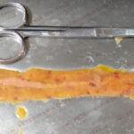
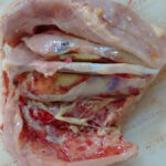
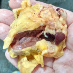
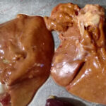
Lesions usually appear in the small intestine – jejunum and ileus –, which shows distention and small necrotic foci observable on the outside walls and on the lumen. The liver can also be affected due to bacteria-produced toxins.


Diagnosis of necrotic enteritis
The presumptive diagnosis is based on:
- The observation of round macroscopic lesions about 2-3 mm in diameter. Concavities are signs of loss of intestinal tissue.
- Gram staining when scraping the intestinal mucosa, where gram-positive bacilli can be observed.
The anaerobic culture of the intestinal content may be of help in the diagnosis. Although it should be noted that the finding of a hemolysis double zone in the culture is not always determinant, since not all strains produce the two toxins responsible for hemolysis. Therefore, it is important to culture the bacteria in a differential medium specifically designed for the isolation of Clostridium perfringens.
The histological study of sick animals shows coagulative necrosis of the intestinal mucosa and masses of bacilli in the fibrino-necrotic debris.
Differential diagnosis
Coccidiosis: Blood in stools is usually associated with an outbreak of coccidiosis or necrotic enteritis, which can usually be differentiated through the macroscopic examination of the lesions observed during the affected animals’ necropsy.
Small intestine enteritis tends to be hemorrhagic in infections caused by Eimeria spp., whereas enteritis caused by Clostridium perfringens tends to be fibrino-necrotic. Coccidiosis rarely produces lesions as acute and severe as necrotic enteritis does. However, both diseases could appear concomitantly.
Mycotoxicosis: Another cause of lesions on the mucosal surface is the presence of trichothecenes – mycotoxins – in the food. These lesions on the mucosal surface tend to be extensive, contrary to the round and well delimited lesions caused by necrotic enteritis.
Treatment and prevention of necrotic enteritis
Antibiotic treatment against necrotic enteritis includes the use of bacitracin, penicillin or lincomycin usually added in drinking water.
It should be noted that dead or dying animals should be quickly removed, since they can be a source of infection due to cannibalism.
Taking prevention measures is of vital importance to avoid a necrotic enteritis outbreak and its subsequent antibiotic treatment. To reduce the incidence of the disease, a first step is to act against its predisposing factors as cited below:
- Reducing the inclusion of Clostridium perfringens-prone ingredients in formulations, such as fishmeal, wheat, barley, rye or oats. In case of not having an alternative – especially when these products must be included in great quantities –, the use of enzymes of non-starch polysaccharides and high-quality proteins would reduce the risk of a disease onset.
- Preventing the action of mycotoxins present in the feed through the use of mycotoxin-binders.
- Preventing digestive parasitosis, such as coccidiosis.
In addition, it necessary to keep a healthy and balanced intestinal flora. Until recent years, antibiotic growth promoters (AGP), such as bacitracin or viriginiamicin, have been used to mitigate the effects of changing intestinal condition. New legal restrictions on the use of antibiotics and growing consumer demands for antibiotic-free products have favored the research of new solutions. Among them, the exclusion of Clostridium perfringens with probiotics and prebiotics, and new additives made of plants.
Compounds
Compounds of botanical origin based on cimenol ring have been proved to have an antimicrobial action – as shown in the test below. They are designed to disintegrate the bacterial cell membrane and penetrate inside, releasing the bacterial content and causing its death.

Conclusions
Necrotic enteritis is an important disease in the poultry industry, due to the economic losses it entails. New regulations and consumer demands for the reduction in the use of Antibiotic Growth Promoters have led to an increase in the appearance of the disease. Current trends are focused on knowing and acting on its predisposing factors.
Bibliography
Lee, K. W.; Lillehoj, H. S.; Jeong, W.; Jeoung H. Y. (2011). Avian necrotic enteritis: Experimental models, host immunity, pathogenesis, risk factors and vaccine development. Poult. Sci., vol 90, 1381-1390.
Paiva, D. & McElroy, A. (2014). Necrotic enteritis: Applications for the poultry industry. The Journal of Applied Poultry Research, Volume 23, Issue 3, 1 September 2014, 557-566.
Skinner, J.; Bauer, S.; Young, V.; Pauling, G. & Wilson, J. (2010). An economic analysis of the impact of subclinical (mild) necrotic enteritis in broiler chicks. Avian Diseases, 54, 1237-1240.
Timbermont, L.; Haesebruck, F.; Ducatelle, R. & Van Immerseel, F. (2011). Necrotic enteritis in broilers: an updated review of the pathogenesis. Avian Pathology, 40, 341-347.
Tsiouris, T. (2016). Poultry management: a useful tool for the control of necrotic enteritis in poultry. Avian Pathol. 2016 Jun; 45(3):323-5.
Wade, B. & Keyburn, A. (2015). The true cost of necrotic enteritis. World Poult. 31. 16-17.


Leave a Reply
You must be logged in to post a comment.