


Mycoses in Poultry
What is mycoses in poultry and how to prevent it?

The mycoses in poultry is caused by fungi, microscopic eukaryotic organisms, which include moulds and yeasts. There is a wide variety of fungal infections in birds, being the most relevant in poultry farming the aspergillosis, candidiasis and mucormycosis. Unlike mammals, dermatophytoses are not common in poultry.
This article will review the symptoms and lesions, diagnosis and control of the most frequent avian mycoses in poultry farming: aspergillosis, candidiasis and mucormycosis.
-
Economic importance of mycoses in poultry
Fungal infections have a great impact on poultry production due to a direct effect on different organic systems and an indirect effect mediated by mycotoxins (toxins produced by fungi). In addition, certain fungi have a zoonotic character, becoming a public health issue, although this type of disease is not frequent in human medicine.
Fungal infections have a lower prevalence than bacterial or viral infections, although they can also present a high mortality and morbidity, especially in young animals. In addition, the lower development of prevention and control methods for fungi compared to bacteria and viruses, results in significant productive and economic losses caused by mycoses in poultry.
-
Transmission and predisposing factors of mycoses in poultry
Fungi are ubiquitous organisms naturally present in the environment. They are usually saprophytic organisms, which tend to multiply in the external environment, mainly in feed and litter, producing spores that act as a source of infection. Mycoses are mostly non-contagious infectious diseases where horizontal transmission lacks of importance.
Most birds are constantly exposed to fungi without any effect, because mycoses in birds are usually related to immunosuppression. Therefore, despite pathogenic fungi are widespread in the environment, immunocompetent birds are generally resistant to the disease. Factors which may cause immunosuppression are:
- Stress situations.
- Environmental conditions (Table 1): High temperatures or high relative humidity, poor ventilation.
- Inadequate farm management: Overcrowding, bad maintenance of the litter or the feed, or prolonged antibiotic therapies. The abusive use of antibiotics favours an imbalance of the intestinal flora that allows the multiplication of fungi.
- Malnutrition situations.

As shown in the charts, yeasts are the most frequent fungi in all seasons, at any time of the year, especially in summer. The second most frequent fungi found in feed depending on the season are Fusarium in winter and spring, Penicillium and Aspergillus in summer, and the genus Mucor in autumn. This information facilitates the orientation in the type of fungal contamination in feed and the possible pathological processes which animals may suffer during the different seasons of the year.
-
ASPERGILLOSIS
Aspergillosis is caused by fungi of the genus Aspergillus. A. fumigatus is the most frequent specie, followed by A. flavus and A. niger. It is an opportunistic fungus, present in the environment that can be present in the lungs and air sacs of the birds with no effects until immunosuppression triggers the disease. Respiratory mycoses are favoured by the avian respiratory system, since the bird’s air sacs are an ideal environment for the growth of Aspergillus.
3.1. Symptoms and injuries
Initial lesions occur in lungs and air sacs, although they can expand from the respiratory system to any other organ, either by direct contact or by hematogenous route, and then cause a systemic disease.
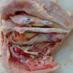
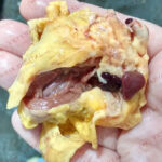
The disease may appear in the acute and the chronic form. Acute aspergillosis usually affects young individuals which do not have a proper development of the immune system, characterized by variable morbidity and high mortality. Animals, usually the ones that are less than two weeks old, have nonspecific symptoms such as lethargy, anorexia and respiratory symptoms, such as dyspnea, gasping, cough and cyanosis. Death of the animal occurs in few days. Affected chicks appear with the beak open, the neck stretched, and the legs slightly apart, in order to get a better breath. Ocular symptoms, such as conjunctivitis and corneal ulcers, may also appear. Lesions that can be observed include thickening of the air sacs membranes, congestive lungs with white mucous exudate, and possible diffuse miliary nodular pattern.
Chronic form of the disease is more frequent and usually appears in adults subjected continuously to predisposing factors. It especially affects turkeys. Respiratory symptoms appear after a long period of disease. Animals may show dyspnea, wasting and, sometimes, nervous symptoms. Less frequently, liver, kidney or gastrointestinal signs may also appear. The most characteristic lesions are sacculitis and white-yellowish granulomas in the parenchyma and serosa of the lungs, although they can also appear in other internal organs.
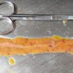
Together with Aspergillus, the genus Fusarium or Penicillium, may cause erosions and ulcers in the corneal layer of the gizzard and can perforate it.
In order to differentiate the origin of gizzard erosions, the corneal layer of the gizzard may be removed to check the back part of this layer. The presence of whitish masses of soft consistency on the back part of the corneal layer confirms the growth of the fungus and, therefore, that it is a mycotic lesion, while the absence of fungal growth confirms that the lesion has a mechanical origin.
3.2. Diagnosis
Antemortem diagnosis of aspergillosis is usually difficult. Post-mortem diagnosis can be made by using swabs of the content in choanae, infraorbital sinus, trachea or air sacs, to perform a cytological evaluation. Tracheal and air sacs swabs are also valid for DNA tests, along with granuloma biopsies.
3.3. Treatment
Different types of antifungals have been tested, although the effectiveness has been normally reduced due to the acute nature of aspergillosis presentation. Furthermore, many practitioners believe that the effectiveness of the treatment does not justify the cost of therapy. It should also be noted that birds that overcome the early stages of the disease will continue to show growth retardation and maintain nodular formations in the thoracic cavity.
-
CANDIDIASIS
This disease is caused by yeasts of the genus Candida, being C. albicans the best-known species. This yeast is part of the digestive flora of birds, and its growth is controlled by the animal’s defensive mechanisms and the bacterial flora. In immunocompromised birds, like young animals or birds subjected to an abusive use of antibiotics, the bacterial flora is affected, and the growth of Candida occurs. This fact may also appear in farms using formaldehyde in feed for Salmonella control.
4.1. Symptoms and injuries
Candidiasis occurs in the gastrointestinal mucosa, especially in the upper and terminal part of the digestive tract, such as the oropharynx, gizzard, esophagus and rectum. Only animals with a severe infection show symptomatology. Birds usually show nonspecific signs, such as anorexia, growth retardation and diarrhea. Infection is visible when located in the oral cavity, seen as whitish plaques under the tongue. Dysphagia may appear if fungal growth is located in the esophagus. The infection can evolve to systemic and show nervous and renal symptoms.
Most common lesions are seen in the upper digestive tract, and consist of thickening of the digestive mucosa, increased mucus and hyperplasia, and whitish multifocal pseudomembranous fibrosis, as well as mucosal ulcers and growth in the oral cavity. Abscesses may also appear in choanae, coming from the growth of the mycelium in the oropharynx.
4.2. Diagnosis
Diagnosis can be made from the identification of the agent in the smear with Gram staining, obtained from swabs or cultures of lesions in the oral cavity or cloaca. Candida lesions must not be mistaken with other lesions of the oral cavity.
4.3. Treatment
Nystatin is the antifungal of choice for the treatment of infections in the digestive tract caused by yeasts, although it is an expensive drug and whose improper use can generate resistance. If the infection is severe or has penetrated the digestive wall, the use of systemic antifungals, such as fluconazole or ketoconazole, will be necessary.
-
MUCORMYCOSIS
Mucormycosis is caused by fungi of the order of the Mucorales, being the most frequent the genera Absidia, Rhizopus and Mucor.
5.1. Symptoms and injuries
Mucorales, which usually come from feed, may spread to different locations, such as the skin, the gastrointestinal tract, the lungs, the eyes or the vertebrae. The excessive use of antibiotics favours their growth.
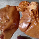
These fungi have special affinity for lymphatic tissue, represented in the intestine by Peyer patches in the duodenum and the ileocecal valve. Due to this, this infection produces an alteration in the immune function of these two types of structures. It is a disease with a long course, in which it can be observed nonspecific digestive symptoms, such as rapid transit, that alternate with periods of no symptoms, which hinders and confuses the presumptive diagnosis.
Necropsy of affected animals may show congestion and inflammation of the ileocecal valve and the Peyer patches.
5.2. Diagnosis
Culture of the agents causing mucormycosis is difficult, as they usually do not grow well in the plaques and the results are not reliable. Histopathology can confirm the presumptive diagnosis. Mucormycosis should be differentiated from other causes of inflammation of the Peyer patches and the ileocecal valve, like the application of highly reactive vaccines.
5.3. Treatment
There is no effective treatment for the disease, although treatment with amphotericin B has been used in humans. Other antifungal treatments, such as nystatin or clotrimazole, have not shown adequate activity in vivo against Mucorales.
-
PREVENTION AND CONTROL OF MYCOSES IN POULTRY
Fungi are opportunistic pathogens; therefore, their control should be done by preventing those factors that cause immunosuppression and favour fungal growth. Thus, it is necessary to check and correct the management and the diet in order:
- Ensure a correct nutrition and adequate feed quality.
- Avoid or minimize stress situations.
- Control the environmental conditions (temperature and humidity) and the litter condition.
- Reduce the use of antibiotics.
In addition, another positive measure that can be applied to reduce the risk of mycosis is based on controlling fungal growth in raw materials by applying preservatives in feed.
There are different types of feed preservatives, such as those based on organic acids, with two months limited effectiveness, and products based on cimenol ring, an active molecule from plant extracts that allows a more efficient and lasting action in the time (up to six months). The molecule is able to drill the fungal and bacterial membranes and alter their cellular metabolism. Cimenol ring has demonstrated microbiocide activity in many in vivo and in vitro trials.

-
Conclusions
Fungal infections have a lower prevalence compared to bacterial or viral infections. However, they constitute an area to be taken into account given their direct and indirect effects on feed quality and animal production. Therefore, it is of great importance to carry out a good preventive strategy through good management practices, adequate nutrition, and the inclusion of preservatives in feed.
Sources:
- Asfaw, M., Dawitt, D. (2017). Review on Mayor Fungal Disease on Poultry. British Journal of Poultry Science, 6(1), pp. 16-25.
- Dalhausen, R. – chapter author (2005). Chapter 29 – Implication of Avian Mycoses in Clinical Disorders. Clinical Avian Medicine – Volume II. Book authors: Harrison, G. J. & Lightfoot
- Refai, M.K.; Osman, G. & Hassan, A. (2016). Monograph on Avian Mycoses & Mycotoxicoses.
- Saif, Y., Fadly, A., Glisson, J., McDougald, L., Nolan, L. and Swayne, D. (2009). Diseases of Poultry. Newark: Wiley.


Leave a Reply
You must be logged in to post a comment.