


Evolution of Cardiac Muscle. Disclosure 8

Myocardial is an organ composed by involuntary striated muscle cells. It is a myogenic muscle, it means self-excited. This characteristic means that it’s started its development at the same time or before the central nervous system therefore its operation is not regulated by it.
In birds and current mammals the heart is tetra cameral, so it has two auricles and two ventricles, separated by muscular walls. The step between auricular and ventricle is through cardiac valves. This myocardial model is the end of a long evolutive way, whose phases can be observed during the steps of the embryological development steps from gastrula to mature being.
So, the first shapes of the heart were simple structures. The most primitive shape existing nowadays can be found in intestinal Ciona (see scientific disclosure 4 (24/09/2010 in Veterinaria Digital). Its heart is formed by one sole dilatation in a blood vessel. The position of this heart is so far from the unique nervous ganglion and without anatomic connexion with it.
Herat of the fishes is more complex with two consecutive cameras, an auricle and a ventricle. This model does not allow still separating arterial from venous blood.
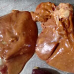
The cardiac evolution leads to a model with two auricles and two ventricles of reptiles and amphibians. Finally the tetra cameral heart appears in birds and mammals.
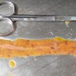
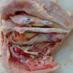
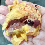
Although the cells of the lefts ventricle and the right ventricle come from two different embryonic nucleus, in essence, the final cardiac muscle behaves a an unique muscle that firstly presented two dilatations and alter formed two more cavities when turned on itself and merged the beginning and the ending. This anatomic structure follows the geometrical models described as Moebius band and Klein surface. These surfaces don’t have interior or exterior and constitute, as the heart, a highly efficient model of mechanic transmission of the force.
The anatomic studies made by Dr. Francisco Torrent, according to which the ventricular part of the heart is a band of muscular continuity, retracting on itself with helix form, during the embryologic development, so the heart is a muscle retracted on itself; and the genomic studies made on intestinal Ciona, allowed to advance in those knowledge and will allow in the future to prevent cardiopathies during embryonic period and improving the efficiency of surgical intervention in the human and animal heart.


Leave a Reply
You must be logged in to post a comment.